MRI for Occipital Neuralgia: A Comprehensive Guide
Table of Contents

Introduction
Are you experiencing persistent headaches that seem to start at the back of your head? You might be dealing with occipital neuralgia, a condition that can be challenging to diagnose. Fortunately, medical imaging techniques like Magnetic Resonance Imaging (MRI) are shedding new light on this painful disorder. In this comprehensive guide, we’ll explore how MRI is revolutionizing the diagnosis and management of occipital neuralgia. Whether you’re a patient seeking answers or a healthcare professional looking to expand your knowledge, this article provides valuable insights into the role of MRI in understanding and treating occipital neuralgia.
Understanding Occipital Neuralgia
Before we dive into the world of MRI, let’s briefly explore what occipital neuralgia is and why it can be so challenging to diagnose and treat.
What is Occipital Neuralgia?
Occipital neuralgia is characterized by pain that starts at the base of the skull and radiates to the back of the head. The pain can feel like a constant, nagging headache that refuses to subside.
Common Symptoms:
- Sharp, shooting pain in the back of the head
- Sensitivity to light
- Tenderness in the scalp
- Pain behind the eyes
- Neck stiffness
The Challenge of Diagnosing Occipital Neuralgia
Diagnosing occipital neuralgia is often tricky because its symptoms overlap with other headache disorders, making it hard to pinpoint the root cause of the pain.
Why Traditional Methods Fall Short
In the past, doctors relied on patient descriptions and physical examinations to diagnose occipital neuralgia. While helpful, these methods don’t always provide enough information about what’s happening inside the body.
Enter MRI: A Game-Changer for Occipital Neuralgia
What is MRI?
Magnetic Resonance Imaging (MRI) is a non-invasive imaging technique that uses powerful magnets and radio waves to create detailed images of the body’s internal structures. Think of it as a super-powered camera that can “see” inside the body.
How MRI Helps in Diagnosing Occipital Neuralgia
MRI is crucial in diagnosing occipital neuralgia because it can reveal underlying issues that contribute to the condition, such as:
- Nerve compression
- Inflammation
- Structural abnormalities
- Vascular issues
The MRI Procedure for Occipital Neuralgia
Before the Scan
Before the MRI, you’ll be asked to remove metal objects to prevent interference with the magnetic field.
During the Scan
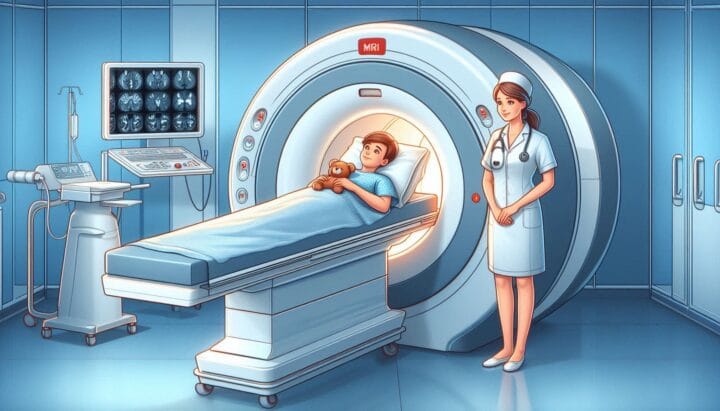
You’ll lie on a table that slides into the MRI machine. It will make loud noises as it captures images, but this is normal.
After the Scan
Once the MRI is complete, you can resume your regular activities. A radiologist will analyze the images and share the findings with your healthcare provider.
What MRI Can Reveal About Occipital Neuralgia
Nerve Visualization
MRI can clearly show the greater and lesser occipital nerves, revealing if they are compressed or inflamed.
Surrounding Structures
Imaging also shows the muscles, blood vessels, and bones around the occipital nerves, helping identify issues that may be contributing to your pain.
Ruling Out Other Conditions
MRI is valuable in ruling out other potential causes of headaches, such as tumors or vascular abnormalities.
Advanced MRI Techniques for Occipital Neuralgia
MR Neurography
MR Neurography is a specialized MRI technique that focuses on imaging nerves. It provides more detailed insights into the condition of the occipital nerves.
Dynamic MRI
Dynamic MRI captures images as you change positions, offering a real-time look at how your neck structures move. This is useful for identifying positional causes of occipital neuralgia.
Interpreting MRI Results for Occipital Neuralgia
What Doctors Look For
Doctors examine MRI images for signs of:
- Nerve thickening or swelling
- Compression points
- Abnormal blood vessel growth
- Structural issues in the neck or skull base
The Importance of Expertise
Interpreting MRI results for occipital neuralgia requires specialized knowledge. Healthcare providers must carefully analyze the images to make informed decisions about your treatment.
How MRI Guides Treatment for Occipital Neuralgia
Targeted Interventions
MRI provides a clear picture of what’s causing your occipital neuralgia, enabling doctors to recommend targeted treatments like:
- Nerve blocks
- Physical therapy
- Medications
Surgical Planning
If surgery is necessary, MRI offers crucial insights to help plan the procedure and increase the chances of success.
The Future of MRI in Occipital Neuralgia Management
Emerging Technologies
New MRI techniques are being developed to offer even more detailed views of nerve function and structure.
Personalized Medicine
As MRI technology advances, it paves the way for personalized treatment plans tailored to the unique anatomical and pathological aspects of each patient’s condition.
Conclusion
MRI has emerged as a powerful tool in the diagnosis and management of occipital neuralgia. By offering detailed images of nerves and surrounding structures, it gives healthcare providers crucial insights to better understand and treat this often-elusive condition. While MRI isn’t always necessary, it’s invaluable in complex cases where traditional methods have fallen short.
As we continue to refine imaging techniques, MRI will likely play an increasingly significant role in helping patients find relief from persistent head and neck pain. If you’re experiencing these symptoms, discussing the possibility of an MRI with your healthcare provider may be the next step toward lasting relief.
FAQs
Q: Is MRI always necessary for diagnosing occipital neuralgia?
A: No, MRI is typically recommended when the diagnosis is unclear or when standard treatments haven’t worked.
Q: Are there any risks associated with MRI for occipital neuralgia?
A: MRI is generally safe, but people with certain metal implants or devices may not be able to undergo the procedure.
Q: How long does an MRI for occipital neuralgia take?
A: Typically, 30-60 minutes, depending on the imaging protocol and use of contrast material.
Q: Can MRI definitively diagnose occipital neuralgia?
A: No, MRI supports the diagnosis but doesn’t always provide definitive answers. Clinical evaluation is also essential.
Q: Will my insurance cover an MRI for occipital neuralgia?
A: Coverage varies by plan, but many insurance providers cover it if deemed medically necessary. Check with your provider for details.
Meta Keywords: MRI for occipital neuralgia, occipital nerve imaging, headache diagnosis, MR neurography, dynamic MRI, nerve compression visualization, occipital neuralgia treatment planning, advanced neuroimaging techniques, personalized headache management, non-invasive pain diagnosis


Post Comment